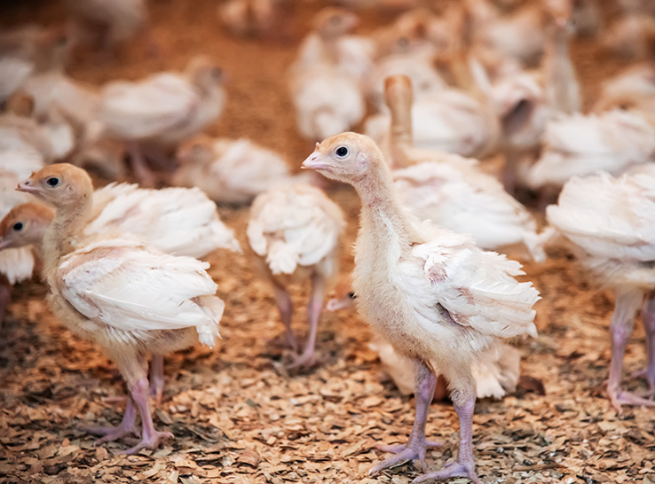

The case of a turkey producer with 10-week-old turkeys experiencing respiratory distress and sudden death proved to be a difficult one to diagnose. The producer sought help from Iowa State University (ISU) Veterinary Diagnostic Laboratory where veterinary student Miranda Painter helped determine the cause of infection.
“This case was in January 2023 and the birds were coughing, had swollen sinuses and were severely underweight,” Painter said. “Given this history, they wanted us to rule out Escherichia coli and check for other diseases.”
Samples were submitted for testing to the diagnostic lab’s bacteriology, PCR and histopathology sections. The results included a few surprises.
“Pasteurella multocida (fowl cholera) came back positive with a CT (cycle threshold) value of 14.8,” Painter said. “Histopathology of the lungs showed diffused areas of edema and hemorrhage. Bacteriology was able to grow E. coli and some staphs.
“Pasteurella isn’t that unusual…and histopathology was able to show pasteurella was present; it had to be the cause,” she said.
Second case
In February, the same turkey finisher contacted the diagnostic lab again for help. A flock of 13-week-old turkeys were having difficulty breathing, coughing and swollen sinuses. There were a few mortalities.
More testing was needed. This time eight turkey heads, livers and spleens along with four oral pharyngeal swabs were sent to the diagnostic lab’s bacteriology and PCR sections. The samples were to be tested for Mycoplasma gallisepticum (MG), fowl cholera, avian influenza, Bordetella avium, Ornithobacterium rhinotracheale (ORT) and Paramyxovirus.
The results were positive for MG with CT values of 29.3 and 36.4, which is what Painter originally thought was the culprit. In addition, bacteriology was able to culture several opportunistic pathogens including E. coli, a strep, several staph infections and ORT.
“The original case was positive for fowl cholera,” Painter said. “So, those samples were retested for MG and came back positive with CT values of 32.3. The second case was positive for MG, so we had the previous samples retested for fowl cholera. They were positive, and the values were 18.4 and 28.2.”
Coinfections confounded diagnosis
“We were able to determine that this flock was dealing with a coinfection of the two diseases — MG and fowl cholera,” Painter explained. “The signs mirrored those commonly found in MG cases with coughing and swollen sinuses but not the signs we commonly see with fowl cholera, like high mortality.”
In addition, there were other opportunistic pathogens identified like ORT and E. coli, which could cover up signs of the cholera and allow the signs of MG to come through, she added.
The takeaway with this case is “don’t be too confident in your gross diagnosis,” she said. “The signs were there that it had to be MG, but for the longest time the results were not coming back to support it.
“In addition, don’t rule out or rule in a disease until you have evidence to back it up,” she continued. “The evidence was there in the first case that it was fowl cholera. But the signs weren’t, so it was ruled out as a false positive until histopathology showed it and the samples were retested.”